About MRI
Magnetic resonance imaging (MRI) is a medical imaging technique used in radiology to form pictures of the anatomy and the physiological processes of the body. MRI scanners use strong magnetic fields, magnetic field gradients, and radio waves to generate images of the organs in the body
The scan is a painless procedure that lasts between 15 to 90 minutes, depending on the size of the area being scanned and the number of images taken.
How to prepare for a MRI scan >

What is MRI for?
MRI scan of the body is mostly performed to evaluate:
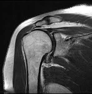
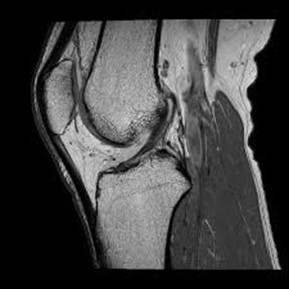
- conditions involving musculoskeletal system including the knee, shoulder, ankle, wrist and elbow as well as ligament, meniscus and rotator cuff tears
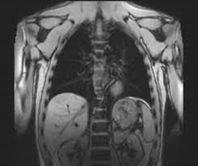
- organs of the chest and abdomen—including the heart, liver, biliary tract, kidneys, spleen, bowel, pancreas, and adrenal glands.
- pelvic organs including the bladder and the reproductive organs such as the uterus and ovaries in females and the prostate gland in males.
- blood vessels (including MR Angiography).
- lymph nodes.
Physicians use an MR examination to help diagnose or monitor treatment for conditions such as:
- injuries, tumors, and degenerative disorders of bone joints, spine, ligaments, meniscus in the skeletal muscular structure of the body
- tumors of the chest, abdomen, or pelvis.
- diseases of the liver, such as cirrhosis, and abnormalities of the bile ducts and pancreas.
- inflammatory bowel disease such as Crohn’s disease and ulcerative colitis.
- heart problems, such as congenital heart disease.
- malformations of the blood vessels and inflammation of the vessels (vasculitis).
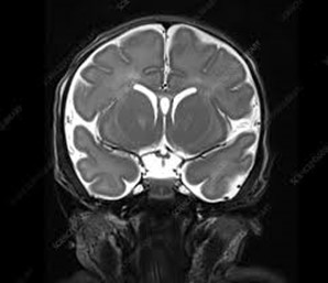
- a foetus in the womb of a pregnant woman.